


Art of Neuroscience Awards 2020
We select some of our favorite entries from this year’s edition of this fascinating competition
The Art of Neuroscience competition started in 2011 at the Netherlands Institute for Neuroscience to make the work of neuroscience more accessible and, if possible, more tangible. The scientists get to evaluate their work and research from a different perspective, and artists are also encouraged to submit work inspired by the brain, creating a cross-over between neuroscience and art.
Here are some fascinating examples of where science meets art inside the most uncharted areas of the human body.
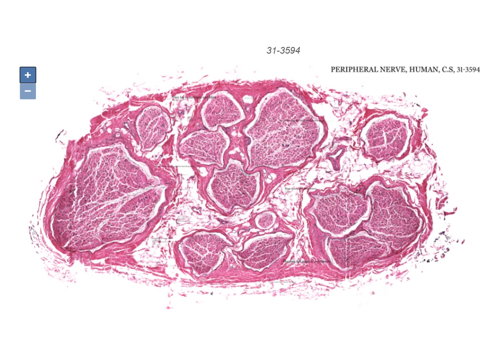
31-3594, Lidija Kononenko, Royal Academy of Arts, London
2020 Winner
What can a picture of a cross-section of a nerve tell us about falling asleep?
This exciting concept is how Lidija Kononenko, a student from the Royal Academy of Arts in London, won the 10th edition of the Art of Neuroscience competition with her artwork ‘31-3594’. In it, she explores the nervous system interactively and playfully. As the page is loaded, you can barely see a pink speck. With each click on the tiny speck, a scan of a peripheral nerve is revealed, accompanied by a text-based story. The artwork is a microscope specimen, a map of symptoms, and an investigation of the unknown.
The interactive work can be viewed here.
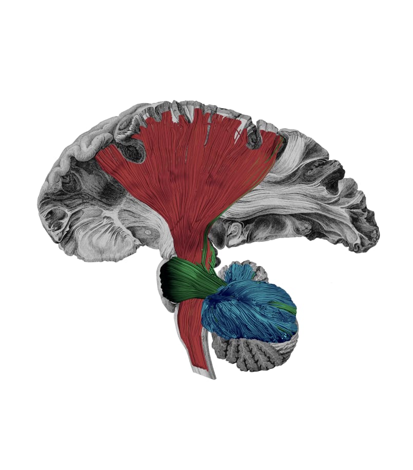
Motor White Matter Networks of the Human Brain, Sanja Budisavljevic, University of St. Andrews, Scotland, 2020 Honorable Mention
In this piece, MRI scans are combined with traditional 19th-century anatomical drawings. The image shows human white matter pathways (important for sensory-motor processing) in color, superimposed on a shaded black and white drawing based on engravings of post-mortem dissections made by scientists in the early 19th century.
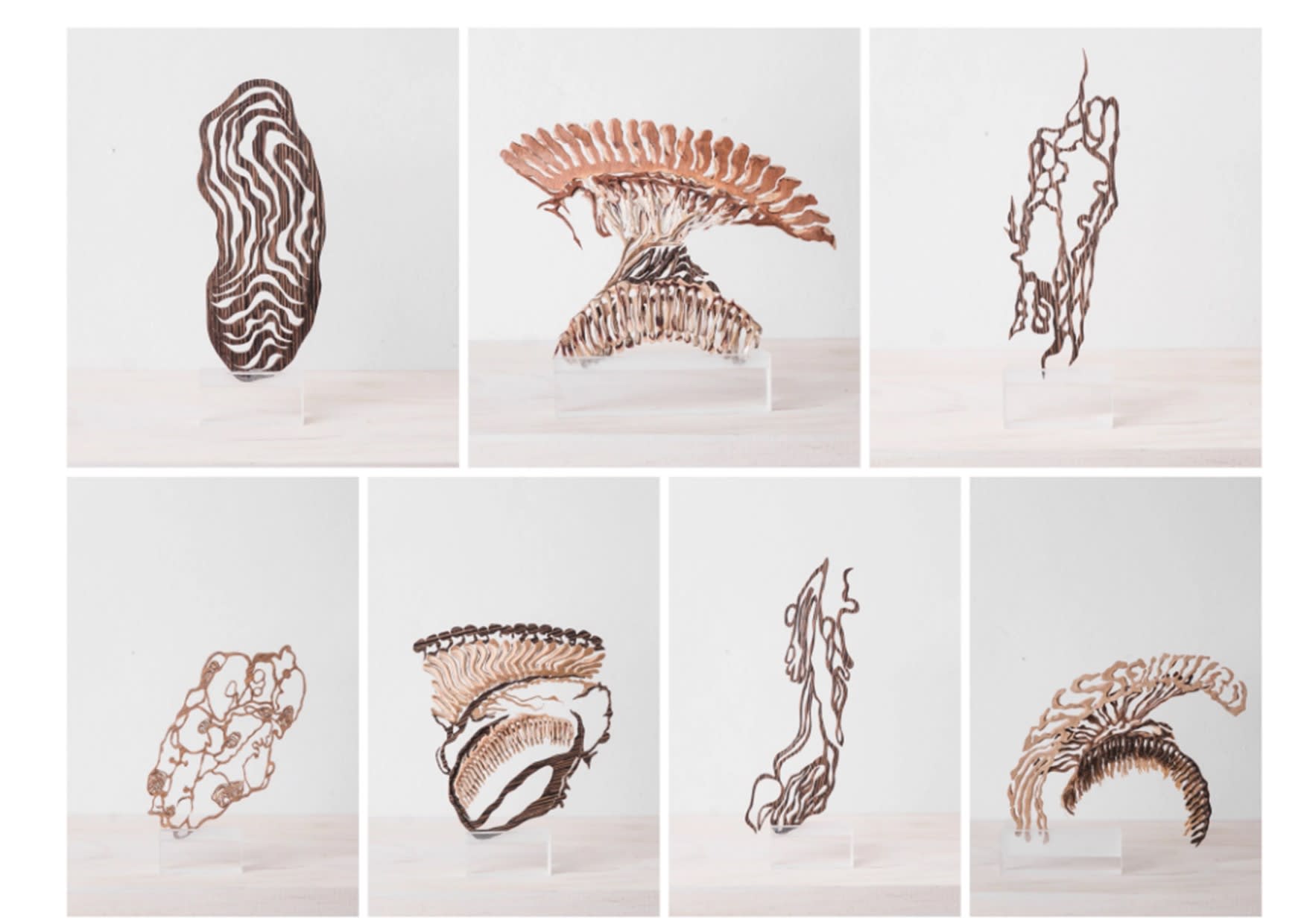
Bdl, Pamela Simard, Montréal, Quebec, 2020 Honorable Mention
The beautiful installations created by the artist remind us of alien organic shapes. They are, in fact, real configurations of synaptic activity (connections between nerve cells in the brain) in the nervous system of an insect, in this case, a fruit fly. Each individual piece was made by hand.
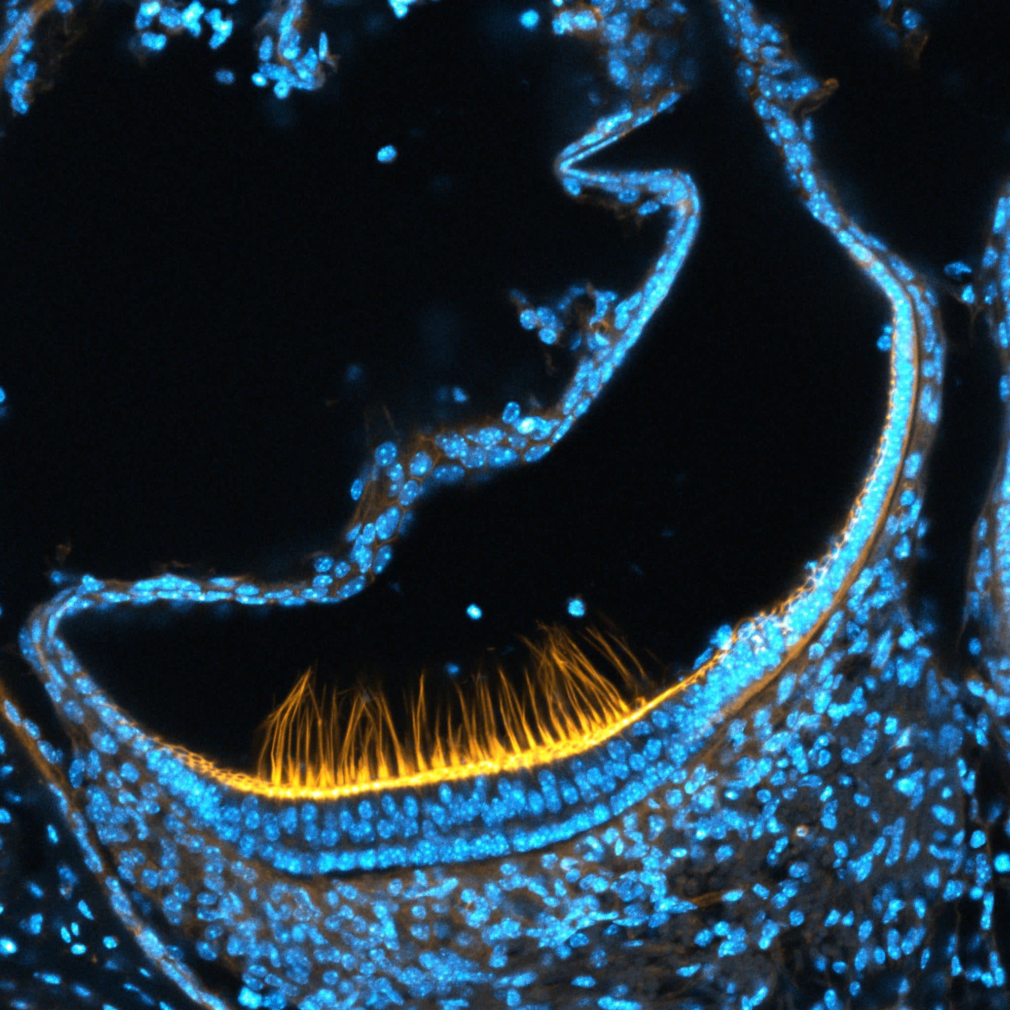
Sensing Spin, Dan Jagger, University College, London, UK
This image captures a fascinating physiological activity in the inner ear. Using a high-resolution microscope, Jagger shows mechanosensory hair cells that are involved in the sense of balance. The yellow in the image represents a protein that helps the tiny hairs stand upright and detect movement as the human head turns.
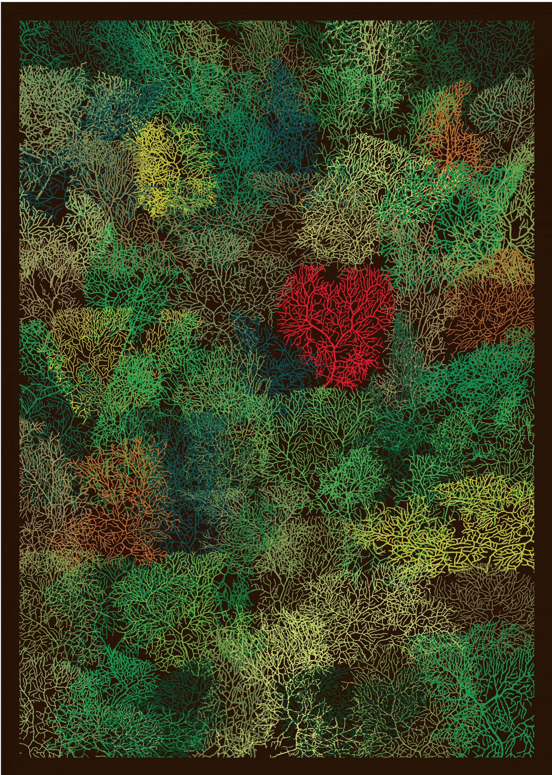
The Protection of Nature Starts in Our Mind, Robert Luck, Heidelberg University, Germany
In this picture, neuroscientist Robert Luck included 65 individually traced images of mice neurons. Luck used the image to raise awareness about climate change and deforestation as it resembles the aerial view of a forest. The number 65 is symbolic as it represents the number of years needed for the rainforest to regrow and gain back 80% of its diversity, and as well as a human lifetime.
More submissions can be found on the Art of Neuroscience website.
You may also like:
- 5 Aerial Photography Instagram Accounts You Should Follow
- Top 10: V&A Illustration Awards
- 20 Museums You Can Visit Online


0 comments